An ependymal cell is a type of glial cell that forms the ependyma, a thin membrane that lines the ventricles of the brain and the central column of the spinal cord. Their main function is to secrete, circulate, and maintain homeostasis of the cerebrospinal fluid that fills the ventricles of the central nervous system. In doing so, they play a vital role in supporting healthy neuronal function and protecting the brain and spinal cord.
What is an Ependymal Cell?
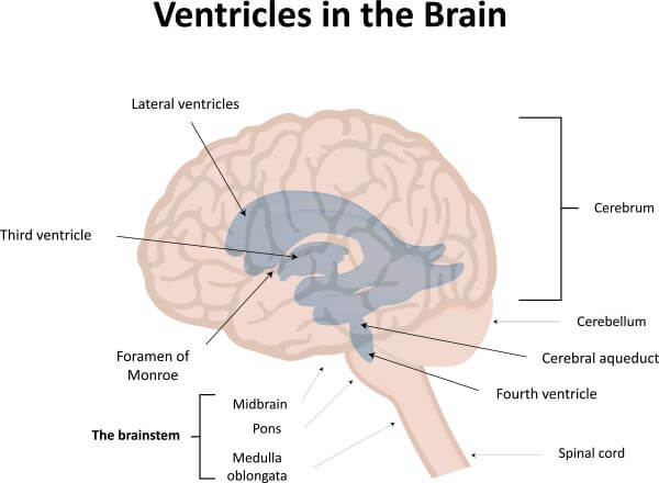
Ependymal cells are one of the four types of glial cells found in the central nervous system (CNS). Collectively, they form the ependyma which is a thin membrane that lines the cavities (or ventricles) in the brain and the central column of the spinal cord. The main role of ependymal cells is to produce the cerebrospinal fluid (CSF) that fills these ventricles.
Where are Ependymal Cells Found?
Ependymal cells line the ventricles of the brain and the central column of the spinal cord. Together they form a thin membrane called the ependyma, which is made of a single layer of ependymal cells and has several functions in supporting healthy neurological function.
Functions of Ependymal Cells
Ependymal cells have several supportive functions to promote healthy neuronal activity. Most of these center around the production of cerebrospinal fluid and the maintenance of homeostasis in the CNS, both of which are essential for healthy brain function.
Production of Cerebrospinal fluid (CSF)
The main function of the ependymal cells is to produce the CSF that fills the cavities of the brain and spinal cord.
CSF has many important roles in protecting and supporting the brain. It acts as a shock absorber and cushions the brain in the event of a blow to the skull. CSF also makes the brain and spinal cord buoyant, which reduces their effective weight and provides further protection against injury.
The CSF also provides nutrients to the brain, and assists in the removal of waste products. By maintaining the environment around the brain, the CSF plays a vital role in supporting and maintaining healthy brain function.
Circulation of Cerebrospinal Fluid (CSF)
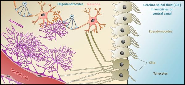
Ependymal cells use cilia (short, motile, hair-like projections on the cell surface) to circulate CSF throughout the ventricles of the CNS. The coordinated ‘wafting’ of these cilia facilitates the flow of CSF from one ventricle to the next, and finally to a region of the brain called the subarachnoid space where is it absorbed. The circulation of CSF is vital for its continued production.
Absorption of Cerebrospinal Fluid (CSF)
Ependymal cells are covered with short, non-motile projections called microvilli. These increase the surface area of the ependymal cells and allows them to reabsorb CSF.
Ependymal Cells and Water Transport
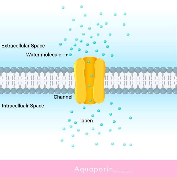
Ependymal cells have water channels called aquaporins which are used to transport water in both directions between the blood and ventricles. This is essential for the formation of CSF, and for maintaining homeostasis of the CSF.
The Ependymal Cell Barrier
The ependyma forms a physical barrier between the CSF and the interstitial fluid (the fluid found between the cells of the spinal cord and brain). This somewhat limits the movement of large molecules between these two regions of the CNS and helps to maintain homeostasis of the CSF and interstitial fluid.
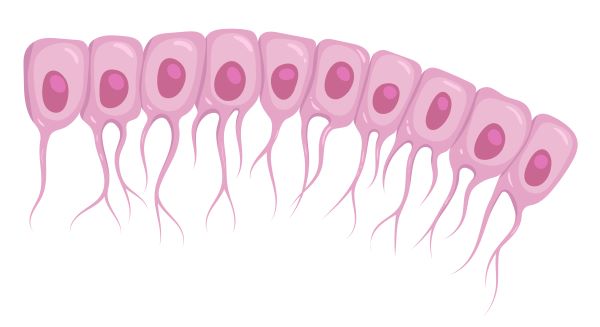
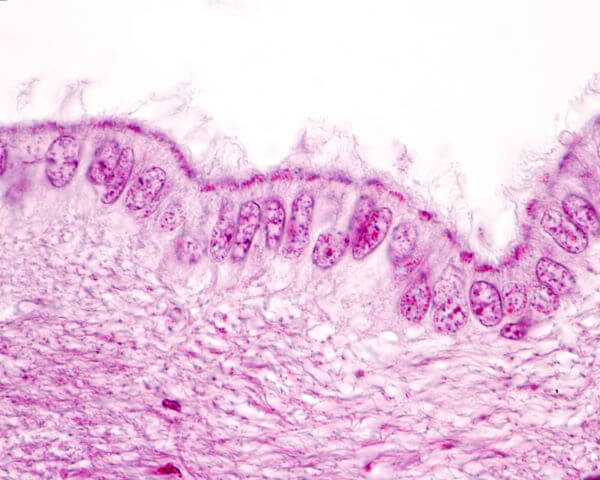
Structure of an Ependymal Cell
Ependymal cells are cuboidal, meaning they are shaped like cubes. On the side of the ependyma that is exposed to the CSF, ependymal cells have several motile, hair-like projections called cilia. They use these to move the CSF through the ventricles of the brain and the central column of the spinal cord. Ependymal cells also have a number of microvilli. These are non-motile structures that increase the surface area of the cell and assist with the reabsorption of CSF.
Other Types of Glial Cells
Ependymal cells are one of the four types of glial cells found in the CNS. Glial cells are one of the two major cell types that make up the nervous system and have a variety of supportive roles in neuronal function. The three other types of glial cells in the CNS are oligodendrocytes, astrocytes, and microglia.
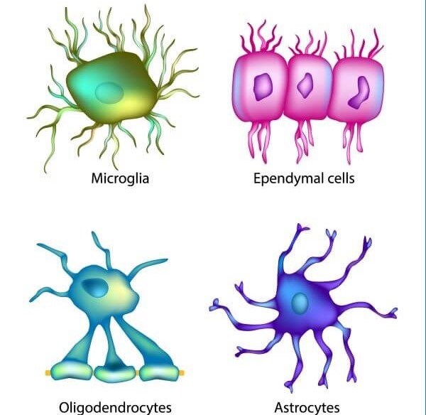
Oligodendrocytes
Oligodendrocytes produce a fatty substance called myelin, which forms an insulating layer around neurons called the myelin sheath. The myelin sheath wraps around the axons of neurons and supports the rapid conduction of nerve impulses along the length of the cell.
Astrocytes
Astrocytes are star-shaped glial cells whose main function is to maintain the chemical environment around neurons. They are the most abundant type of glial cell in the brain and assist in regulating blood flow, providing nutrients to nerve tissue, and recycling waste products in the brain.
Microglia
Microglial cells are specialized immune cells of the CNS and support healthy brain function by removing damaged neurons and infectious agents.