Definition
The cranial nerves of reptiles, birds, and mammals consist of twelve pairs of numbered peripheral nerves that originate in the cerebrum and brainstem and have their own specific sensory and motor pathways. Lower vertebrates such as fish and amphibians only have ten pairs. Cranial nerves are motor, sensory, or mixed neurons that bring motor and sensory messages to and from the face, neck, and shoulders, as well as many internal organs such as the heart, lungs, and gastrointestinal tract.
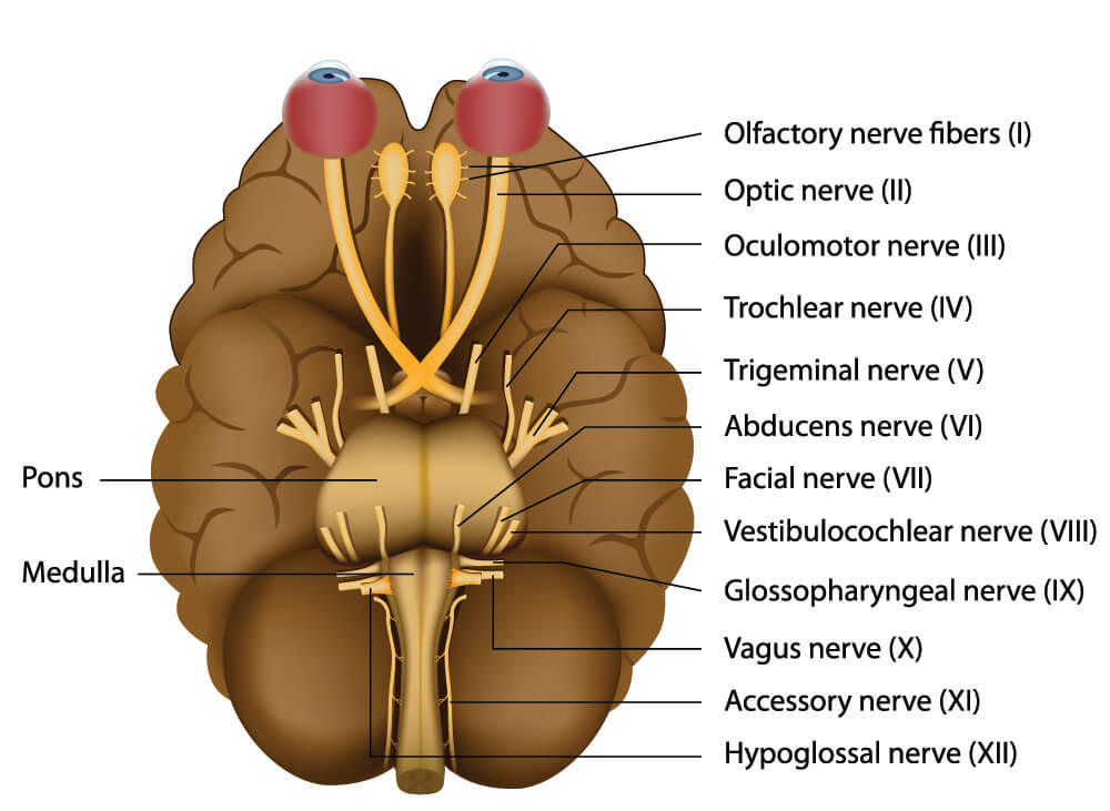
Cranial Nerve Function
Cranial nerve function depends on whether each nerve is composed of motor, sensory or mixed nerves, and also on the region where the nerve endings are located. Motor nerves leading away from the brain (efferent nerves) to the muscles and sensory nerves that bring information to the brain (afferent nerves) provide signals that enable movement, organ function, and sensations such as pain, smell, taste, vision, hearing, and touch. Mixed nerves, as their name suggests, have both sensory and motor nerve pathways. When studying the cranial nerves, it is easier to look at the different functions of each nerve pair.
Cranial Nerve I Function
The olfactory nerve, or the first cranial nerve, is a purely sensory nerve pair (afferent nerves). They enable the sense of olfaction or the ability to smell. Our brain responds to tiny vapor bubbles made of gaseous odors. The olfactory nerve is very connected to other sensory nerves such as those that supply information about taste. While taste and smell are not the same, together they help us to attribute food with flavor. Even if you have lost your sense of taste you might still be able to detect distinct flavors – most of the flavor of the food you eat comes from your sense of smell, provided by the olfactory nerve.
Cranial nerve I is also very associated with the emotion and memory areas of the brain. That fresh cut-grass smell that makes you think of summers at home? That is a result of earlier cranial nerve I information in combination with your memory and emotions.
The olfactory nerve is the shortest cranial nerve and is composed of special visceral afferent nerve cells or SVAs. SVA neurons can sense stimuli in the internal organs and mucous membranes and work closely together with the gustatory (taste) processes of the brain.

Our sense of smell begins with vaporized odor molecules that enter the nostrils and dissolve into the moist layer of mucus. Just under this mucus, we find the olfactory epithelium. This layer of tissue contains olfactory sensory neurons and odor receptors. Both the olfactory nerve and our sense of taste share these receptors. When the odor receptors are stimulated, electrical messages are sent to the olfactory bulb at the back of the nose. The olfactory bulb forwards information to a part of the brain that processes the information, but it also sends signals to the limbic system (emotion and memory) and the neocortex (conscious thought).
Cranial Nerve II Function
The optic nerve or cranial nerve II is another purely sensory nerve with special somatic afferent (SSA) neurons that bring vision, hearing, and balance information to the brain. The rod and cone cells of the retina pick up different light wavelengths and send electrical stimuli via the retinal ganglia to the optic nerve. The optic nerve ends at the optic chiasm of the brain where the nerve fibers cross, bringing information from the right eye to the left cerebral hemisphere of the brain and vice versa.
Cranial Nerve III Function
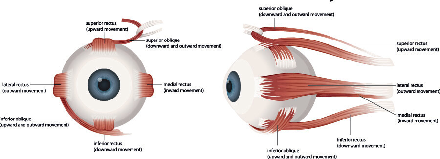
The oculomotor nerve is responsible for the movements of the pupil and lens of the eye, the upper eyelid, and visual tracking and gaze fixation muscles. It is primarily an efferent motor nerve consisting of general somatic efferent (GSE) neurons. GSEs bring impulses from the central nervous system to skeletal muscles. They allow us to close our eyes or move our eyes when looking around us.
Cranial nerve III also has general visceral efferent (GVE) neurons that innervate smooth muscle. Smooth muscle contractions are autonomous (involuntary) functions. This means we never have to think about adjusting the size of our pupils or making the lens fatter or thinner.
The oculomotor nerve has a minor sensory function, conveying afferent sensory information from selective muscles of the eye as part of the pupillary light reflex. This part of cranial nerve II detects the amount of light coming in through the pupils. The information it provides will be used to influence the GVE neurons that adjust pupil size.
Cranial Nerve IV Function
The trochlear nerve or fourth cranial nerve also controls eye movement. It is a somatic efferent motor nerve for voluntary skeletal muscle movement. In this case, the muscle is the superior oblique muscle that allows eye rotation and the act of looking down (to look at where this muscle is located, you will have to look up…at the eye muscle diagram). This is a pulley system that prevents the eye from rolling towards the back of the head.
Cranial Nerve V Function
Cranial nerve five or the trigeminal nerve innervates the face. It is a mixed motor and sensory nerve. This cranial nerve contains special visceral efferent (SVE) neurons that conduct impulses to the skeletal muscles of the face, jaw, and neck via three branches. The first branch (V1) is the ophthalmic nerve, the second (V2) is the maxillary nerve, and the third (V3) is the mandibular nerve. Using general somatic sensory nerves, CN V also provides sensory information from the face. Cranial nerve V has no autonomic (involuntary) fibers but travels alongside the involuntary nerves of other mixed cranial nerves that regulate the many glands of the face and neck.
Cranial Nerve VI Function
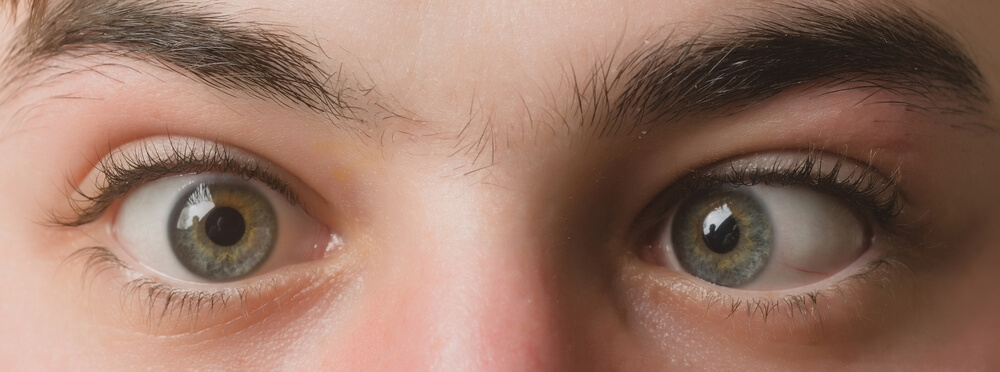
The abducens nerve or cranial nerve six is a purely efferent (motor) nerve that runs to the ipsilateral (same side of the body) lateral rectus muscle of the eye orbit. This muscle is at the outer side of the eyeball (see above image). It allows for eye abduction or movement away from the midline. You can remember this by thinking about the other use of this verb. To abduct someone is to take them away; however, unlike the victim, this is not involuntary movement.
Several syndromes including Foville’s, Millard-Gubler, and Raymond’s syndromes all present with sixth nerve paresis symptoms. Sixth nerve paresis, often referred to as sixth nerve palsy, causes one or both eyes to turn towards the nose due to (partial) paralysis of the lateral rectus muscle.
Cranial Nerve VII Function
The facial nerve or cranial nerve VII is a mixed nerve. Motor neurons control the muscles of facial expression, part of the digastric and stylohyoid muscles under the jaw, the stapedius (ear bone muscle), and the occipitofrontalis (forehead muscle). Sensory neurons transmit taste sensation from part of the tongue as well as from the external auditory canal via special visceral afferent (SVA) nerve fibers. Finally, general visceral efferent nerves innervate the submandibular, sublingual, and lacrimal glands of the face, as well as the mucous membranes of the nose and mouth.
Cranial Nerve VIII Function
The function of cranial nerve VIII or the vestibulocochlear nerve is purely sensory. It helps us to perceive the sounds in our environment and our position and movement within it. These stimuli are processed in our auditory (hearing) and vestibular (balance) systems. This nerve is composed of two types of nerve fibers – vestibular and cochlear.
The cochlear nerve brings sound stimuli originating from the cochlea to the brain. It is responsible for our perception of balance (equilibrium), specifically the position of the head in relation to the rest of the body. The vestibular nerve transmits input from the semicircular canal of the ear. All of these functions use special somatic afferent (SSA) nerve fibers.
Cranial Nerve IX Function
Cranial nerve IX or the glossopharyngeal nerve is composed of motor and sensory nerve fibers and is a mixed nerve. Like the oculomotor (lens and pupil), facial (glands), and vagus (various organs and emotions) cranial nerves, it also plays a role in the parasympathetic nervous system via general visceral efferent (GVE) fibers. This parasympathetic function regulates the secretions of the parotid glands, our largest salivary glands.
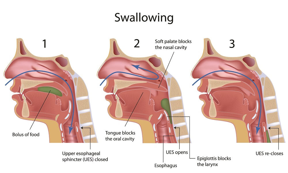
Via special visceral efferent fibers, our pharynx lifts voluntarily whenever we speak or swallow, a function known as the branchial motor component. The gag reflex is another example of this efferent function; another is the swallowing reflex – the image below shows the various steps of this involuntary action. Some older people with significant nerve degeneration where CN IX is damaged or less effective have a much higher risk of choking as both the coordination and the response time of the swallowing reflex are affected.
Sensory or afferent nerves produce three other cranial nerve IX functions. General visceral afferent nerve fibers bring sensory information from baroreceptors and chemoreceptors in major arteries so we can automatically respond to changes in blood pressure, and blood oxygen and carbon dioxide levels. General somatic afferent fibers bring sensory information from the inside of the tympanic membrane, upper pharynx, and the back of the tongue to the brain. Finally, special visceral afferent nerve fibers forward signals from the taste buds located at the back of the tongue.
Cranial Nerve X Function
Another mixed nerve, cranial nerve X or the vagus nerve, is most commonly associated with fainting; however, it has many more roles than this. The term vasovagal syncope only helps us to remember one of them.
This longest cranial nerve runs through the head, neck, thorax, and abdomen. Due to its size, the vagus nerve is usually split into four sections – the cranial, cervical, thoracic, and abdominal divisions. It has a parasympathetic function, meaning it fires impulses when we are at rest. CN X encourages peristalsis of the colon when we are relaxing or sleeping via general visceral efferent fibers. These fibers are also responsible for the involuntary muscle control of the heart, lungs, and esophagus.
Special visceral efferent nerve fibers provide movement of the muscles of the throat. Special visceral afferent fibers bring sensory information in the form of taste from the epiglottis, and general somatic afferents allow this same sensation in the soft palate, pharynx, and larynx mucosae. Finally, general visceral afferent innervation allows the stretch receptors (baroreceptors) and chemoreceptors of the large arteries, gathering information together with CN IX.
Vasovagal syncope is fainting in the presence of certain triggers. These can range from the sight of blood to being surprised, or from standing up too rapidly to having a bowel movement. Vasovagal syncope occurs when the vagus nerve is overstimulated. Overstimulation of the parasympathetic reflex means the vagus nerve slows the heart rate and dilates the blood vessels causing a rapid drop in blood pressure. Less oxygen reaches the brain and we fall out of consciousness.
Cranial Nerve XI Function
The function of cranial nerve XI, otherwise known as the accessory nerve or spinal accessory nerve is a motor nerve responsible for voluntary (somatic) innervation of the muscles of the shoulder, neck, and throat. It is the only cranial nerve that also has neurons in the spinal cord.
The cranial part of the accessory nerve or cranial component is responsible for muscle control in the larynx, pharynx, and soft palate of the throat. The spinal component enables trapezius and sternocleidomastoid movement – whenever you shrug your shoulders you are making use of CN XI.
Cranial Nerve XII Function
The last of the twelve cranial nerves is the hypoglossal nerve – a motor nerve that focuses on the muscles of the tongue.
The tongue has eight muscles that are either intrinsic or extrinsic. The four intrinsic muscles are not attached to bone. They allow the tongue to change shape. These muscles work to lengthen, shorten, curl, and flatten the tongue. The four extrinsic muscles are attached to bone and are responsible for tongue position. They retract, trough, and elevate the back of the tongue respectively.
List of 12 Cranial Nerves
This list of twelve cranial nerves includes basic, comparative information for easy reference. S stands for sensory and M for motor function.
I Olfactory S – Olfaction
II Optic S – Vision
III Oculomotor M – Eye muscles
IV Trochlear M – Superior oblique (eye)
V Trigeminal S – Facial sensation M – Mastication
VI Abducens M – Lateral rectus (eye)
VII Facial S – Taste, hearing M – Facial expression, glands
VIII Vestibulocochlear S – Equilibrium, hearing
IX Glossopharyngeal S – Taste, carotids M – Swallowing, saliva
X Vagus S – Visceral sensation M – Parasympathetic
XI Accessory M – Neck, shoulders
XII Hypoglossal S – Taste M – Tongue
Quiz