Definition
Cartilage is a semi-rigid but flexible avascular connective tissue found at various sites within the body. With a pliable structure composed primarily of water, this tissue type is also extremely tough. Cartilage is found throughout the human body in areas such as the joints, nose, airway, intervertebral discs of the spine, and the ear.
Cartilage Function
Cartilage function is more than structural, and has different functions in the life cycle. In the embryo, it provides support and is a precursor to bone. Embryonic cartilage either remains as cartilage or provides a substructure for endochondral ossification, meaning it also functions as a template for the rapid growth and development of the musculoskeletal system.
Cartilage is a supple tissue which allows for facial movement as well as providing a lightweight supportive structure in the external ear, and the tip and septum of the nose. In other regions it acts as a shock absorber, cushioning areas where bone meets bone and preventing abrasion and damage. A joint would also not be able to bend without the flexibility of cartilage. A combination of roles is seen in the airways, where cartilage rings around the trachea prevent collapse and damage, and cartilage at the ends of the ribs allows the ribcage to swing upwards and outwards during inspiration. Cartilage also plays a role in bone repair where, as in the embryo, it provides a template for ossification, this time to broken sections of bone.
Types of Cartilage
There are three cartilage types in the human body. Although their components are very similar, the quantities of each component differ, providing different qualities to each type. Accordingly, each type has a particular location.
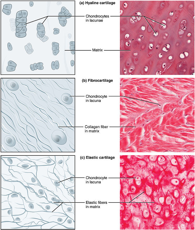
Hyaline Cartilage
The most common form of cartilage is hyaline cartilage. Hyalos is the Greek word for glass, which describes the appearance of this type of connective tissue – translucent, blueish-white, and shiny. Hyaline cartilage is usually only 2 – 4 mm thick (all cartilage must be thin, as there is no vascularization in this tissue type, and nutrients and oxygen must be obtained through diffusion). It is the embryonic form of cartilage, and also found in the ribs, joints, nose, larynx and trachea.
Hyaline cartilage collagen fibers are primarily type II, extremely thin, and invisible to the microscope due to similar refractory properties to that of the matrix itself.
Fibrocartilage
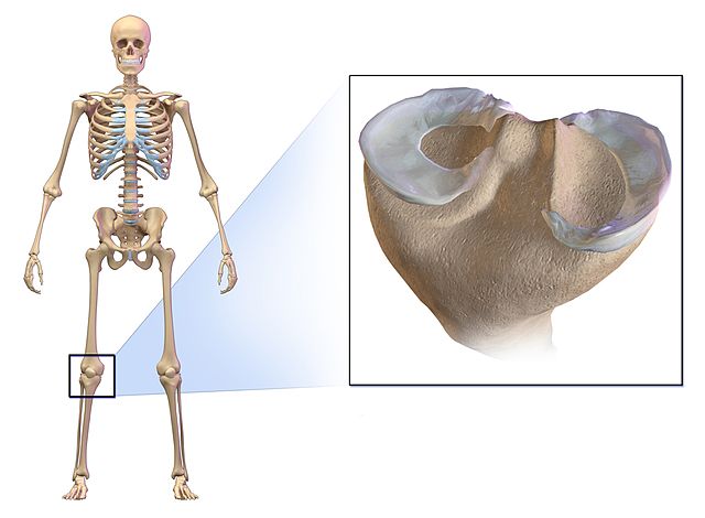
Found where tendons and ligaments meet bone, at the pubic symphysis, in the menisci, the sternoclavicular joint, and the annulus fibrosus (the center of the intervertebral disc), fibrocartilage is a very strong and pliable connective tissue. It is reinforced with collagen fiber bundles that run parallel to each other, allowing a low level of stretch. Because of the abundance of collagen fibers, fibrocartilage is white in appearance. It lacks a perichondrium and is composed of type II and type I collagen fibers. The image below shows the smooth, white horseshoe shape of the fibrocartilaginous menisci.
Elastic Cartilage
Elastic cartilage is primarily found in the external ear (auricle or pinna), the Eustachian tube, and the epiglottis. These parts of the anatomy are required to always spring back into the original shape. Elastic cartilage’s role is purely structural, offering flexibility and resilience due to a mixture of elastic fibers and type II collagen fibers. It is yellow in color, and without the organized structure of fibrocartilage when viewed on a microscope slide.
The Main Ingredients of Cartilage
Cartilage is made up of highly specialized cells called chondrocytes and chondroblasts (chondro refers to cartilage), and other extracellular material which forms the cartilage matrix.
All connective tissue types within the human body are derived from the embryonal mesoderm. Bone, the strongest of the connective tissues, is the last to form and can remain in cartilage form well after birth. Increased cartilage to bone ratio enables a flexible and pliable new-born to exit the birth canal. A new-born has 300 bones, as opposed to the 206 of the normal adult, and all of these originate from cartilage.
From the seventh week of embryonic life, the process of ossification or osteogenesis slowly replaces cartilage with bone. This process continues into early childhood. Cartilage grows in two ways. In interstitial growth, chondrocytes proliferate and divide, producing more matrix inside existing cartilage throughout childhood and adolescence. In appositional growth, fresh layers of matrix are added to existing matrix surface by chondroblasts in the perichondrium. The perichondrium is a dense layer of connective tissue which surrounds most cartilage sites. Its outer layer contains collagen-producing fibroblasts, while the inner layer houses large numbers of differentiated fibroblasts called chondroblasts.
Chondroblasts
As long as they are free to move, chondroblasts produce the elements of the extracellular matrix (ECM). This cell type first forms a matrix of hyaluronic acid, chondroitin sulphate, collagen fibers, and water during embryonal development. Chondroblasts eventually become immobile after becoming surrounded by the matrix, and are then referred to as chondrocytes.
Chondrocytes
Chondrocytes are the immobile form of chondroblasts. They are surrounded by the matrix and contained within allotted spaces called lacunae. A single lacuna can contain one or more chondrocytes. Chondrocytes have varying roles according to the type of cartilage they are found in. In articular cartilage, found in the joints, chondrocytes increase joint articulation. At growth plates, chondrocytes regulate epiphyseal plate growth. While chondroblasts are ECM manufacturers, chondrocytes maintain the existing ECM and are a less active form of the same cell.
Fibroblasts
Fibroblasts are found in all types of connective tissue. In cartilage, these cells produce type I collagen. In certain situations, fibroblasts transform into chondrocytes.
Extracellular Matrix
There is significantly more matrix than cells in cartilage structure, as the low oxygen environment and lack of vasculature do not allow for larger numbers. Because of this, there is little metabolic activity, and little to no new growth in cartilage tissue – one of the reasons the elderly commonly suffer from degenerative joint pain. Cartilage does continue to grow slowly, however. This can be seen in the larger ears and noses of older individuals.
The ECM of cartilage contains three characteristic elements:
Collagen
A protein-based collagen matrix gives form and strength to cartilage tissue through a mesh-like structure of fibrils. Although there are many different forms of collagen in the human body, the collagen found in cartilage is primarily type II, with an attached FACIT (short for fibril-associated collagen with interrupted triple helix) XIV collagen which determines the diameter of these fibers.
Proteoglycans
Proteoglycans are large molecules that bind with water, providing flexibility and cushioning qualities. Proteoglycan monomers bond to hyaluronic acid by way of link proteins, as is the case with the large proteoglycan Aggrecan (chondroitin sulphate proteoglycan 1), seen below.
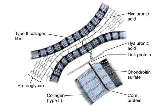
The high numbers of negative charges such constructions provide, together with a large surface area, make it possible for proteoglycans to bind to large amounts of water. This creates high osmotic pressure, increases load-bearing, and constitutes the gel-like consistency of the ECM.
Noncollagenous Proteins
Noncollagenous elements of the ECM are small in number and supposed to play a role in maintenance and organization of the cartilage structure on a macromolecular level.
Quiz