Definition
Adipose tissue is split into two main types of connective tissue – white and brown – that store and burn energy respectively. White adipose tissue also provide a layer of insulation, while brown adipose is found in too small quantities (in children and adults) to do this. Brown fat does, however, release energy in the form of heat. Adipose tissue is made up of adipocytes – differentiated cells that store excess energy as triglyceride droplets, together with various supporting cells and fibers. Fat cells also have an endocrine function as they can secrete hormones.

Adipose Tissue Function
Adipose tissue function depends on the type and location of fat within the body. Brown and white fat are found in all warm-blooded and cold-blooded animals. It was formerly thought that birds do not have brown fat, but this has since been disproved. As adipose tissue function depends primarily on the fat type, it is better to look at brown and white fat function separately, although some characteristics overlap.
White Adipose Tissue Function
White adipose tissue functions as a storage and insulating layer under the skin but also plays an endocrinological role in the body. Too much fat produces more chemicals but also increases the risk that the body gradually stops responding to these chemicals as well as usual. That means that metabolic disorders such as diabetes, and inflammatory diseases are more likely to develop over time if we are overweight. Fat is so important to homeostasis (stable body processes) that it is now considered to be a fully-fledged organ rather than connective tissue with an energy storage function.
White adipose tissue (WAT) has a number of functions, depending on where it is found in the body. These include angiogenesis (the production of new blood vessels) and blood coagulation (clotting), reproduction, glucose metabolism, fat metabolism, the regulation of appetite, immunity, and vascular tone or how much a blood vessel can contract and dilate.
There are two sub-types of WAT: subcutaneous WAT (sWAT) and visceral WAT (vWAT). Areas are divided into depots, for example the perigonadal (around the gonads) and retroperitoneal (behind the peritoneum) depots. Where these depots are located can mean differences in cell type, their distribution, and their form, as well as differences in function according to which adipose tissue genes are expressed. Visceral WAT – white fat that gathers around the organs – has been linked to metabolic disorders. If you have an ‘apple’ shape, you have a greater percentage of visceral fat than someone with a ‘pear’ shape. This is considered less healthy. A pear shape is the result of larger deposits of subcutaneous WAT and may, in contrast to the apple, be a protective characteristic.
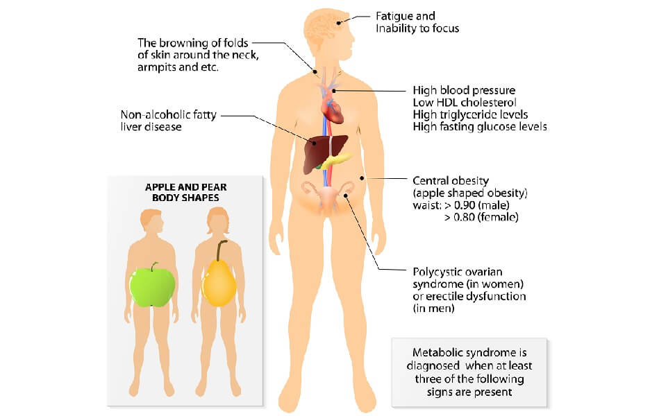
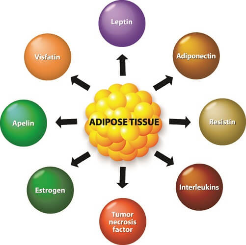
White adipose tissue cells have large vacuoles and low numbers of mitochondria. They have the ability (and the space) to store lipids in the form of triglyceride droplets. This stops these fatty acids from circulating in the blood which can cause inflammation of the blood vessels and arterial plaque build-up. However, white adipose tissue is not simply a fat reservoir. WAT is an endocrine organ that secretes hormones, growth factors, enzymes, matrix proteins that form protein fibers, and cytokines (immune response) – the most important can be seen in the image below. Different chemicals are produced at different locations depending on which adipose cell genes are switched on or expressed. For example, more leptin (appetite suppression) and adiponectin (glucose and fatty acid regulator) are produced in sWAT, and more interleukin-6 (inflammatory response) and plasminogen activator inhibitor 1 (blood clotting) are produced in vWAT. These chemicals (adipokines) can be pro-inflammatory or anti-inflammatory and ratios seem to be unbalanced in obese individuals. Other chemicals, when produced in excess or insufficient amounts, produce the symptoms of metabolic syndrome – high blood pressure, apple shape, insulin resistance, and high cholesterol and/or triglyceride levels in the blood. You can see the signs in the above image. Up to one-third of US adults suffer from metabolic syndrome.
Brown Adipose Tissue Function
Brown adipose tissue or BAT was previously thought only to have a heat-generating role but we now know that it also produces various adipokines. When brown fat was transplanted into test animals in the laboratory, scientists saw that their glucose tolerance and insulin sensitivity improved. Brown fat transplants or administering the chemicals they produce may be a future treatment for diabetes, metabolic syndrome, and even obesity.
There is also a subcategory of brown fat called beige adipose tissue that lies interspersed within brown adipose tissue. Beige fat is thought to be especially important for the treatment of metabolic disorders. This is because the highest numbers of growth factors, hormones, and cytokines are produced in beige fat cells.
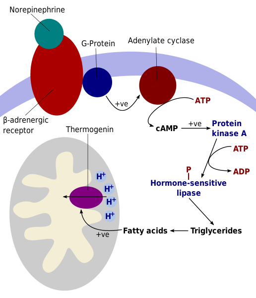
Heat production in BAT involves the large numbers of mitochondria in brown adipocytes as well as a central protein called Uncoupling Protein 1 (UCP1) or thermogenin. When we feel cold, our central sympathetic nervous system releases norepinephrine. Norepinephrine tells the mitochondria to produce heat. Other chemicals are necessary, for example, thyroid hormone is required for BAT cells to respond to norepinephrine. People with untreated hypothyroidism where the thyroid gland does not produce enough of this hormone often feel cold, whatever the environment.
In this heat-generating response, UCP 1 – the picture below labels it thermogenin – the purple oval in the gray mitochondria – is released from the mitochondria of the brown adipose tissue. This protein reduces adenosine triphosphate (ATP) production. ATP is the result of the conversion of oxygen and nutrients into usable energy – a process known as cell respiration. Further on in this article you will find a detailed diagram of intracellular energy production.
By stopping cell respiration, heat builds up within the fat cells. You might think that by not producing as much ATP the body does not require as much energy. This is not true. Generating heat requires significant energy, about four times more than the energy released by working muscle tissue. But that is not a problem when you are not significantly underweight, as this extra energy is immediately available in the triglyceride-filled vacuoles of each fat cell. If you happen to be in a cold environment for a long period of time, these triglycerides become depleted. You then need to make energy from chylomicrons (fat and protein globules) in the intestinal tract, lipoproteins stored in the liver, and circulating glucose. This is still, efficiency-wise, preferable to using the lipids found in white tissue adipocytes. As many of us know, white fat can be very tough to get rid of!
The body’s other mechanism for heat production – shivering – does not happen as frequently in people who have higher levels of brown fat. New-born babies, for example, do not shiver. Only at around six months of age (when their levels of brown fat have significantly reduced) does the shiver reflex begin in humans. That is why the production of heat by brown fat cells is called non-shivering thermogenesis.
Adipose Tissue Location
Adipose tissue location changes as we age. While newborns have very little WAT, this is the predominant type in adults. Subcutaneous WAT is located under the skin and above the muscle in an area called the panniculus adiposus. Women tend to have more sWAT in the thighs and breasts; men have more abdominal sWAT. Visceral white adipose tissue is found in the omentum, mesentary, and retroperitoneal space, as a covering layer of some internal organs, and in bone marrow.
Brown adipose tissue is found in higher quantities in new-born babies; they have a low proportion of white adipose tissue and this makes them much more susceptible to hypothermia. Without lots of BAT, babies would be in extreme danger in temperatures under 96°F (35.5°C). As we grow older, the ratio of white to brown fat changes; thicker insulating layers of white fat mean there is less need for BAT thermogenesis. In adults, most brown fat is located behind the peritoneum, around the major blood vessels, deep in the neck, between the shoulder blades, and along the back.

Adipose Tissue Structure
Adipose tissue structure is fairly uncomplicated. This tissue consists of large quantities of adipocytes and their precursors (preadipocytes) and some other cell types. All is enclosed within a fibrous extracellular matrix that is very well connected to blood and lymph vessels.
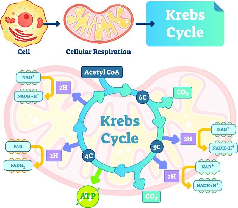
Subcutaneous WAT and visceral WAT are formed from different progenitor cells and do not express the same genes. They are, physiologically speaking, dissimilar, although anatomically they look almost the same. The color is the most distinguishing difference when you don’t have a microscope. All WAT is yellowish due to the large lipid droplets that are contained in a single intracellular cavity (unilocular cells). Visceral WAT contains unilocular but also multilocular cells and these have higher numbers of mitochondria; it looks similar to brown fat in appearance. You can refresh your memory by returning to the first image in this article that shows white, brown, and beige adipocytes. Higher numbers of mitochondria mean more cellular respiration which usually provides energy (see the Krebs cycle image below). This role can be substituted by thermogenesis in brown and beige adipose tissue under the stimulation of the sympathetic nervous system that responds to cold temperatures.
Fat cells need to be in direct contact with a blood supply because they absorb fatty acids through the blood vessel walls. Fats either need to be broken down in the intestinal tract from dietary fats or have to be converted from carbohydrates in the liver in a process called hepatic de novo lipogenesis. When small enough, fatty acids enter the adipocyte cell membrane via passive and active transport mechanisms. All adipocytes contain a range of organelles in the cytoplasm that include mitochondria, Golgi apparatus, endoplasmic reticulum, ribosomes, one or multiple vacuoles, nucleus, and nucleolus. Adipocytes have a stronger membrane than many other cell types – they are similar in strength to bone and cartilage cells.
Adipose tissue contains mainly adipocytes with other cells such as fibroblasts, stem cells, macrophages, T-cells, B-cells, mast cells, eosinophils, neutrophils, and dendritic cells scattered throughout the tissue. The fibrous matrix consists of collagen fibers and through this matrix runs a network of nerve fibers and lymph and blood vessels. Non-adipocytes are grouped under the term stromal vascular fraction, where stromal refers to adipocyte-supporting cells and vascular to the blood supply. There are a lot of white blood cells in adipose tissue – many scientists consider weight-related disorders to be auto-immune disorders.
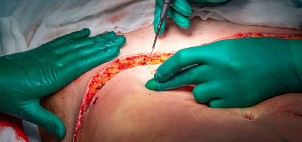
The white fat adipocyte contains a central vacuole that fills with triglycerides, free fatty acids, cholesterol, and simple glycerides. Up to 95% of the body’s lipids can be stored in fat tissue at any one time, the majority in WAT. WAT cells become so full with lipids that the cytoplasm and organelles get squashed against the cell membrane, giving an adipocyte its distinctive round shape. Large numbers of lipid-filled adipocytes in white adipose tissue are easy to see if the skin has a deep enough cut. They are round and yellow, as you can see below.
Brown and beige fat cells are unilocular and multilocular (containing multiple vacuoles) and multilocular cells have much higher numbers of mitochondria; this means they are better at generating heat. The gene that expresses uncoupling protein 1 is highly active in BAT but nearly silent in white adipose tissue. Brown and beige fat tissues also play endocrine roles like white fat and secrete similar adipokines. Beige adipose fat tends to contain larger vacuoles and slightly fewer mitochondria than brown adipocytes, showing that the heat-producing characteristics are primarily the function of brown adipose tissue.