Definition
The choroid plexus is part of the blood-brain barrier that protects the central nervous system from harmful chemicals, and is the primary source of the various components of cerebrospinal fluid. The choroid plexus or plica choroidea borders the membrane of the pia mater and the ventricles of the brain. It is an area of specialized cells that surrounds a direct blood source (capillary). The choroid plexus also plays neuroendocrine, neuro-immune, and excretory roles.
Choroid Plexus Location
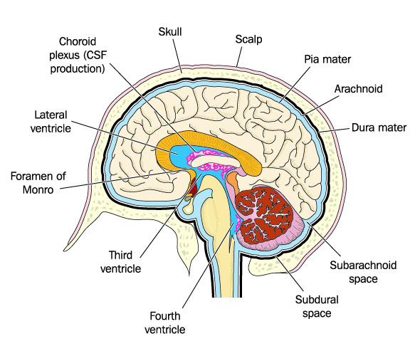
Choroid plexus location is within all four ventricles of the brain, even though many textbooks do not show the presence of the choroid plexus at the fourth ventricle. Choroid comes from the Greek word chorioeides which means skin-like. It should be imagined as a capillary-rich membrane that is spread along part of the inside layer of the ventricles. While the eye choroid is a layer of connective tissue, the choroid plexus of the brain is a layer of epithelial cells, tela choroidea (connective tissue), and many capillaries. It is only found in the central nervous system.
The choroid plexus is part of the blood-cerebrospinal fluid barrier or blood-brain barrier. Its cells produce cerebrospinal fluid at the top of the lateral ventricles, through a space called the foramen of Monro, along the top of the third ventricle, and in a small section of the fourth ventricle.
The choroid plexus is composed of ependymal cells, a type of glia. It is a specialized area within the ventricular membrane.
To better place the location of the choroid plexus, we should look at the surrounding anatomy.
The soft tissue of the brain is surrounded by three membranes called the meninges. These are the outer dura mater, central arachnoid mater, and inner pia mater. The choroid plexus is located within the membranous pia mater, the innermost meninges layer – this membrane is indicated by a black line in the below image. The pia mater lines the ventricles and is attached directly to the brain by astrocytes. Cerebrospinal fluid is excreted from the upper surface of the choroid plexus and flows from the direction of the pia mater up into the subarachnoid space.
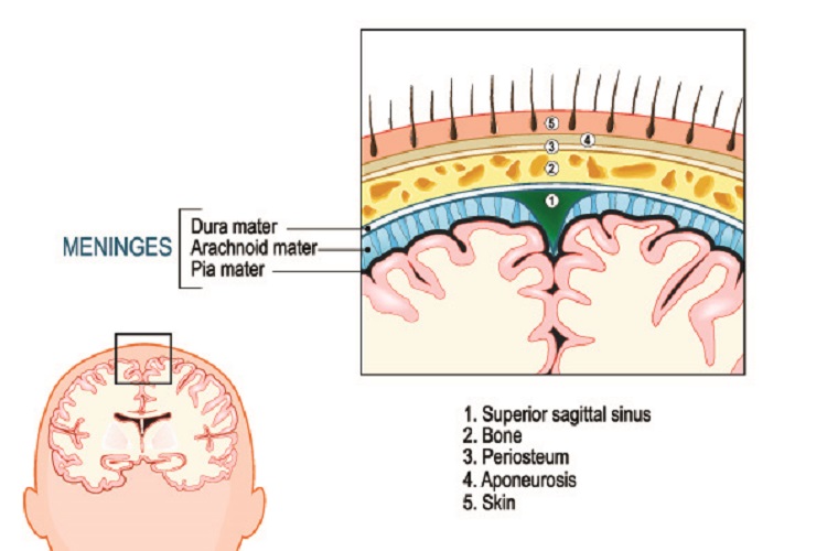
Between the pia mater and arachnoid mater lies the subarachnoid space. Sub means under – the subarachnoid space lies directly under the arachnoid mater. You might recognize the word and think it sounds like the Latin name for a spider – you are correct. The arachnoid is a thin membrane connected to a web-like sheet. It is this connected sheet’s similarity to a spider’s web that gives the arachnoid membrane its name.
The web is formed by arachnoid trabeculae – connective tissue filaments that help to keep the brain in place. The subarachnoid space also houses cisterns – interconnected compartments that more widely separate the two inner meninges and provide space for nerves and blood vessels. If a cistern becomes blocked, the cerebrospinal fluid cannot move to the next compartment and this will increase pressure within the brain.
How much or how little CSF is produced is controlled by the sympathetic and parasympathetic nervous systems. Parasympathetic control reduces the amount of cerebrospinal fluid that is produced in the choroid plexus, while the sympathetic nervous system increases blood flow to this area and so increases CSF production.
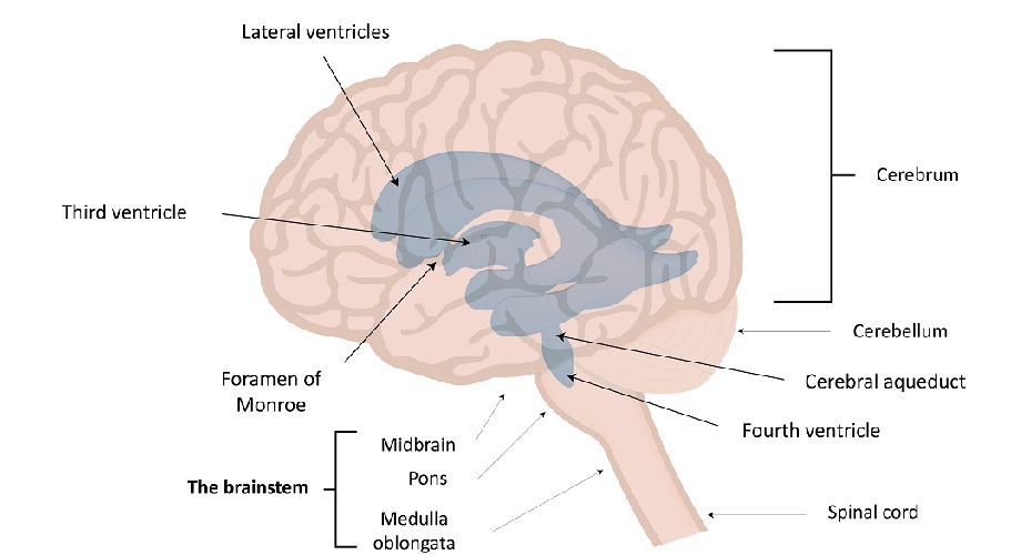
As the choroid plexus is located at the ventricles, knowledge of the brain ventricular system is also helpful. This system is composed of a left and right lateral ventricle at either brain hemisphere connected to a single third ventricle by the foramen of Monro. The third ventricle is connected to the fourth ventricle by the cerebral aqueduct. The fourth ventricle is connected to the spinal cord via two holes – the foramen of Luschke and the foramen of Magendie.
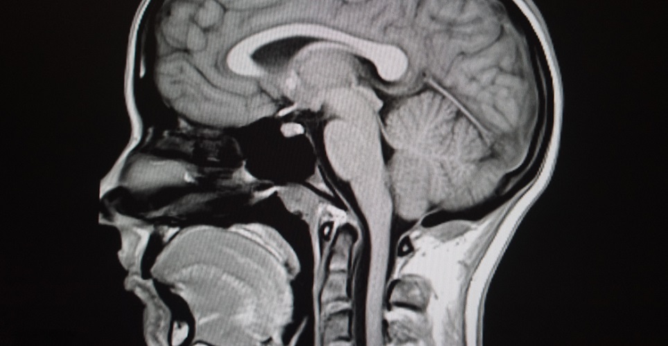
The wishbone shape of each lateral ventricle can be divided into sections. The anterior horn lies at the front of the brain (temporal lobe); the body of the lateral ventricle follows it, splitting into the pointed posterior horn in the occipital lobe and the posterior lobe that follows the shape of the temporal lobe. Both anterior horns end at the foramen of Monro that forms a conduit into the single third ventricle. The MRI image below shows the white curved corpus callosum that sits on top of the lateral ventricles. The black area underneath is due to the fluid within the ventricle. Above the cabbage-like cerebellum is the black region of the third ventricle, and in the center of the cerebellum you can clearly see the triangular outline of the fourth ventricle.
The third ventricle is located between the two arms of the wishbone-form lateral ventricles. The bottom of the third ventricle opens into the aqueduct of Sylvius or the cerebral aqueduct. This channel runs from the third to the fourth ventricle.
It is at the fourth ventricle that CSF exits the brain and enters the subarachnoidal space of the spinal cord. The fluid flows through two channels – the foramen of Magendie and the foramen of Luschka. In this photograph the cerebral aqueduct is indicated with the number nine. The roof of the fourth ventricle that enters the cerebellum is shown by the number eight. This is at the level of the pons (10). The fourth ventricle (11) has two drainage points – the right foramen of Luschka (L) and the midline foramen of Magendie (M).
When picturing the choroid plexus, you should also know how its tissue is arranged. The outermost layer (epithelium) consists of simple columnar ependymal cells (glia cells). These surround projections of pia mater tissue and have central capillaries.
It is these specialized cells, with their many cilia to increase the total surface area, which excrete the products that form cerebrospinal fluid. Ependymal cells in the choroid plexus do not secrete CSF but the individual components of this fluid such as water, ions, oxygen, growth factors, and nutrients. Cerebrospinal fluid is an ultrafiltrate of blood plasma.
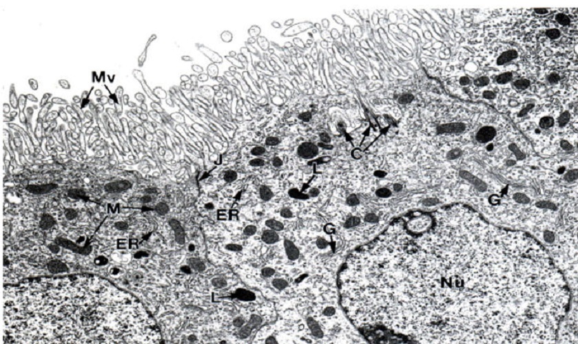
Microvilli also absorb waste products and return them to the blood circulatory system. The image below looks at the choroid plexus epithelium under the microscope. Mv indicates the microvilli, J points to the closed junction between cells that is tightly sealed to provide an efficient blood-brain barrier. C points to centrioles, G to the Golgi apparatus, and ER to the endoplasmic reticulum. The nucleus is labeled Nu. All of the white space above the microvilli (the inside of the ventricles) is full of CSF.
On the inner side of the pia mater a basement membrane gives a point of attachment for the ependymal cells. The highly vascular connective tissue of the pia mater is the source of the choroid capillaries, and every protrusion of this connective tissue houses a capillary. It is important to know that the ependymal cells of the choroid plexus are stuck closely together. In this way, molecules that enter and leave the cerebrospinal fluid are highly controlled. This is the principle of the blood-brain barrier.
Choroid Plexus Function
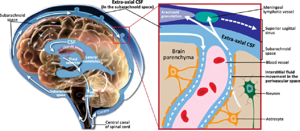
Choroid plexus function is not just the production of cerebrospinal fluid (CSF), although this is a primary role. CSF is made by choroid plexus ependymal cells. This fluid is excreted out of the epithelium into the fluid-filled space of the ventricles. It flows via gravity into the subarachnoid space of the brain and spinal cord, helped by our breathing and arterial pulse that cause gentle turbulence. This turbulence distributes the various components of CSF throughout the subarachnoidal space.
The choroid plexus ependymal cells are covered with microvilli, multiple projections with centrally-located capillaries that are fed by the choroidal and cerebellar arteries. Hair-like structures called cilia on the surface of ependymal cells also help to distribute the components of cerebrospinal fluid.
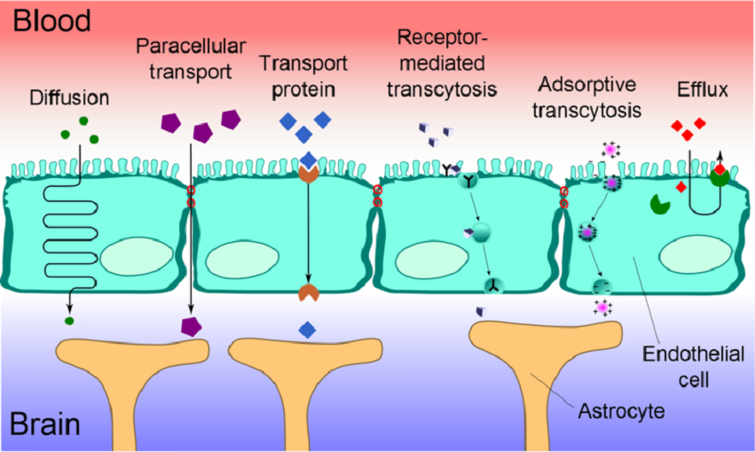
The epithelium of the choroid plexus does not leak in the same way as other epithelial layers. This is very important, as the choroid plexus is part of the blood-brain barrier (BBB) that protects the brain from harmful chemicals. The tight junctions between adjacent cells (red circles in the below image) let very little into the brain and most chemicals require transporter proteins to move through the ependyma. The blood-brain barrier (image below) separates blood plasma from the interstitial fluid of the central nervous system. While many products can get in and out, this barrier is not easily accessible by larger hydrophilic molecules such as amino acids and glucose – these need special transporter proteins.
Although an adult only has approximately 25 ml of CSF in the ventricles at any one time, another 115 to 245 ml flows through the subarachnoid space of the spinal cord. The fluid constantly drains through the lining of the spinal arachnoid membrane and is absorbed into the venous blood circulation and lymph nodes. This means the choroid plexus must produce approximately 650 ml CSF every 24 hours. How quickly CSF is absorbed back into the blood supply depends on the pressure within the subarachnoid space. When the pressure of the cerebrospinal fluid is higher than the venous pressure, it will be absorbed into the arachnoid until these pressures equalize.
More recent research points to multiple additional roles and functions of the choroid plexus. These include choroidal cytokines that globally affect the brain, healing characteristics due to the presence of stem cells, the ability of the choroid plexus to relay hormone-produced signals to the hypothalamus, and its being a possible access point for gut bacteria. This is a two-edged sword, as gut microbiota can both cause damage to the cerebrospinal system or provide immune cells that protect it. It is thought that the choroid plexus can metabolize rather than only absorb toxins, and that it can detect signs of and respond to inflammation. Brain regeneration (plasticity) may also require input from the choroid plexus.
The ependymal cells of the choroid plexus can secrete large volumes as the microvilli provide an optimal surface area for countless transport proteins. If all of the microvilli were stretched flat, the surface area of the choroid plexus would be around five square meters. Ependymal cells have high numbers of mitochondria that provide the energy for these many active transport channels.

When the blood-brain barrier is not working as it should, the central nervous system is open to infection. One of the signs of multiple sclerosis is the formation of plaque on the inside of the ventricles caused by pathogenic white blood cells that would normally not be able to enter the barrier. The blood-brain barrier prevents many medications from reaching the brain and much pharmacological research is underway to find drugs that can reach the central nervous system.
Cerebrospinal Fluid
Cerebrospinal fluid sometimes referred to as liquor, is primarily produced in the choroid plexus. It is an alkaline, clear liquid composed of 99.13% water. CSF allows the brain to ‘float’ – the average adult brain weighs around 1500 grams, but by surrounding it with a layer of fluid the effective weight is only around fifty grams. This means that if the head is slightly moved, shaken hard, or hit, the brain does not crash against the skull with as much force, limiting damage.
CSF is relatively similar in composition to blood plasma in terms of molecule types, but the concentrations of these molecules substantially differ. As an ultrafiltrate of blood plasma, liquor contains various nutrients and metabolic products that support the surrounding tissues. It, therefore, contributes to homeostasis. Samples of CSF can be collected with a lumbar puncture using a hollow needle inserted into the subarachnoid space. In the photograph, the second red injection site is where a local anesthetic has been injected as this technique is relatively painful. You can see a clear drop of CSF at the collector tip.

The following products are present in CSF:
- Ions
- Sodium
- Potassium
- Calcium
- Chloride
- Nutrients
- Glucose
- Lactate
- Lipids
- Immunoglobulins and other small proteins
- Urea
- White blood cells (occasionally)
Too low volumes of cerebrospinal fluid inside the ventricular system are most commonly due to leakage or ependymal damage. Aging may also affect how much CSF is produced as the ependymal cells work less efficiently.
High volumes are caused by sympathetic nervous system disorders that encourage the ependymal cells to produce high levels of CSF. Other causes are infection and choroid plexus cancer. Some causes are idiopathic (unknown).
A further function of cerebrospinal fluid is its role as a waste-disposal system. There is no lymphatic system in the brain; this means that CSF also contains metabolic waste products. The ependymal cells that allow the components of CSF to enter the ventricular space also remove waste products.
Choroid Cyst
A choroid cyst can form during fetal growth when a small fluid-filled bubble becomes separated during the anatomical development of the choroid plexus. This can happen in up to 2% of the population and does not necessarily cause an effect. The bubble can be detected during a gestational ultrasound and is then referred to as a choroid cyst. In the ultrasound image below the cyst is marked between the two crosses.
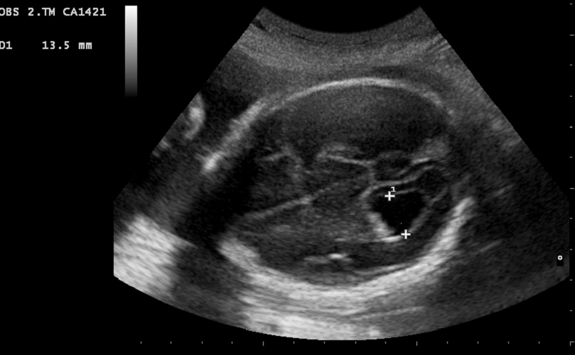
Trisomy 18 or Edwards syndrome is a chromosomal condition associated with various physical and physiological abnormalities and early death. One sign that is nearly always present in fetuses with trisomy 18 is the choroid cyst. The cyst of a fetus is rarely cause for concern on its own but is an indication of this chromosomal disorder.
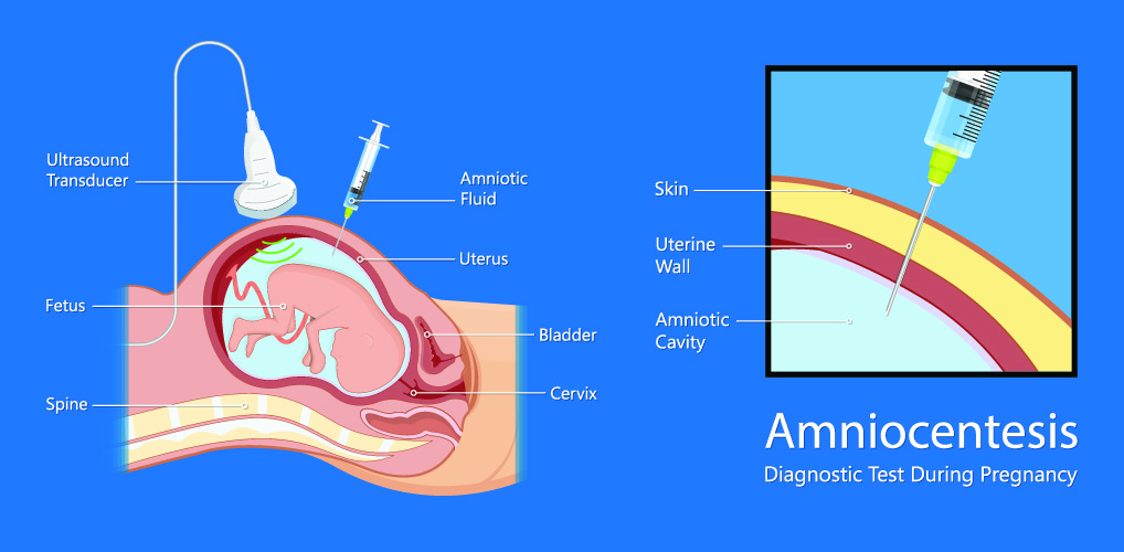
On a prenatal ultrasound, choroid cysts can be detected as spaces of at least two to three millimeters in diameter. In most cases, these are no reason for concern but parents may be advised to undergo an amniocentesis to test for trisomy 18. The association between a choroid cyst and a severe chromosomal defect has led to often unnecessary anxiety. While a choroid plexus cyst is found in around one in one hundred fetuses, trisomy 18 is much rarer – around one in 3,000.
Some research associates autism to choroid cysts, although results are mixed. The presence of multiple subarachnoid pseudocysts – cysts that are not surrounded by ependymal cells – have been found in children that later developed autism and attention deficit hyperactivity disorder (ADHD). The presence of a choroid cyst, however, does not mean a child will present with either of these disorders.
Other types of choroid cyst can form throughout life. These can be benign or malignant (cancer). Any malignancy in the brain should be treated where possible. Benign choroid cysts only require treatment if they contain infected material (pus), are large, or begin to grow larger and cause higher pressure within the ventricles. Other terms used to describe a choroid cyst are arachnoid or leptomeningeal cyst. A larger choroid cyst can push into the arachnoid as the gap between the pia mater and arachnoid is very small.
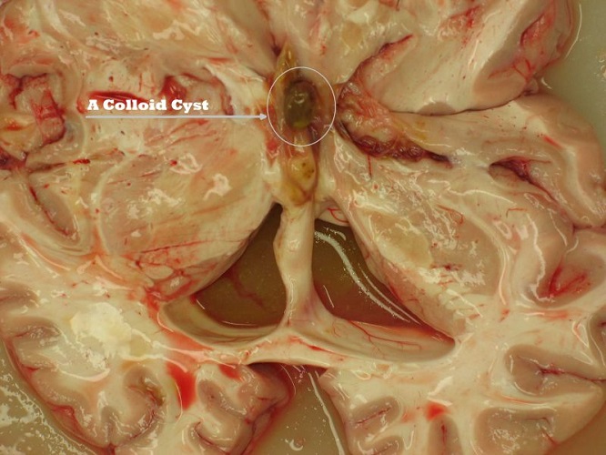
A colloid cyst is a cyst within the fluid of the ventricle – within the CSF. Colloid refers to a gel-like blob that may or may not affect the flow of cerebrospinal fluid. A colloid cyst is not a choroid cyst. This colloid cyst had become lodged inside the third ventricle.
Choroid cyst symptoms involve a build-up of pressure. If the ependymal cells of the choroid plexus are constantly producing cerebrospinal fluid in all four ventricles, a build-up of fluid will occur above any full or partial blockage. If a sink drain is plugged with hair and the sink is full, it will empty at a much slower rate as water finds its way through the individual hairs. However, if you leave the tap on (ependymal cells are always ‘on’), the sink will overflow.
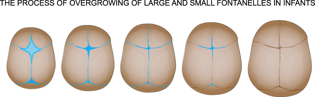
As the brain is covered by a solid, immobile skull (from the age of 10 to 24 months of age), there is no overflow. The ventricle above the blockage expands and this expansion pushes onto the brain.
In the very young, the skull is flexible – the different bony plates have not yet fused. If the ventricles swell, the brain is pushed against the flexible skull. Over time, the skull grows to accommodate the increased pressure. A child with untreated hydrocephalus (water on the brain) will develop a significantly-sized skull but will not have as many other symptoms caused by high pressure as the skull’s expansion protects the brain. In adults or in children where a blockage is severe and the skull is not given time to adjust, common symptoms are headaches, nausea and vomiting, feelings of dizziness, problems seeing or hearing, pain in the face, and imbalance.
Choroid Cancer
Choroid plexus cancer grades can be found on the National Cancer Institute website. This form of cancer most commonly occurs in young children. They may be caused by certain genes but the cause is usually unknown. Unlike most choroid cysts, chemotherapy, radiotherapy and/or surgery are required. The blood-brain barrier makes chemotherapy treatment less effective.
As with large choroid cysts that push into the ventricular space, rare choroid plexus tumors can create increased intracranial pressure (ICP) with the associated symptoms. As the aqueducts of the ventricular system are particularly narrow, even small tumors can cause severe blockages. The pressure within the ventricles can be measured by inserting a catheter into the brain. This catheter can also drain fluid away, reducing any pressure.
Choroid Plexus Fissure
A choroid plexus fissure (choroidal fissure) is a natural anatomical structure. It is at this point that the choroid plexus attaches to either lateral ventricle. Only the choroid plexus and arachnoid lie between the lateral ventricle and the choroidal fissure. Cysts can also form here – usually benign choroidal fissure cysts. This part of the lateral ventricle is usually used as a point of navigation for neurosurgeons and radiologists.
Hydrocephalus
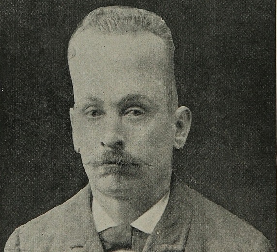
Hydrocephalus is a relatively common occurrence in children; it has been reported in various ancient texts. In adults, acute hydrocephalus is not physically visible as the fused skull is unable to accommodate increased pressure in the brain – diagnosis is usually based on symptoms such as headaches, imbalance, nausea, and visual disturbances. In children, the skull will gradually expand to relieve the pressure caused by excess cerebrospinal fluid production or ventricle blockage. This man from the early twentieth century had suffered from hydrocephalus when very young. The cause would most probably have been non-obstructive hydrocephalus.
Hydrocephalus that is not caused by obstruction occurs in approximately one in 1,000 births. It is usually the result of subarachnoid bleeding or infection, or the presence of a choroid plexus papilloma that makes the ependymal cells produce much higher quantities of CSF.
Hydrocephalus due to obstruction is called obstructive or non-communicating hydrocephalus. The ependymal cells above the blockage continue to produce CSF but this fluid cannot properly drain into the subarachnoid space of the spinal cord. The ventricle(s), aqueduct(s) or foramen(s) above the blockage will swell. This swelling pushes the affected ventricles outwards and creates pressure within the skull.
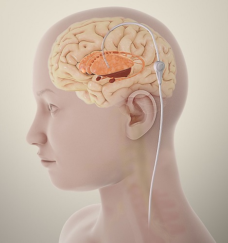
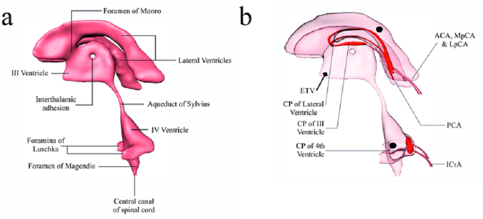
Hydrocephalus treatment requires a shunt that creates another route for the CSF above the blockage. Usually, these shunts – long, flexible tubes – open into the peritoneal space of the abdomen. Where the cause is an overproduction of CSF in the ependymal cells, it is possible to burn away some of these cells and so lower the produced quantities. Endoscopic third ventriculostomy is relatively new technique used to treat obstructive hydrocephalus caused by blockages in the aqueduct between the third and fourth ventricles.
A shunt is placed through the membrane of the floor of the third ventricle into the roof of the fourth ventricle, bypassing the cerebral aqueduct. The point of perforation can be seen in the right-hand image (ETV). The choroid plexus at the lateral, third, and fourth ventricles is depicted in red. The blood supply is indicated in dark pink
Quiz